Cell migration plays a fundamental role in veins and arteries development, i.e. vascular development and angiogenesis. Despite cell movement being first seen more than 300 years ago, the mechanisms and identification of the influencing factors behind cell movement are an experimental challenge. In this project, we combined a microfluidic platform that reproduces chemotactic cell migration through the extracellular matrix and a biophysical modelisation through phase-field modelling.
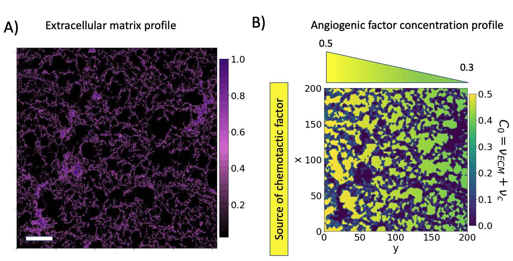
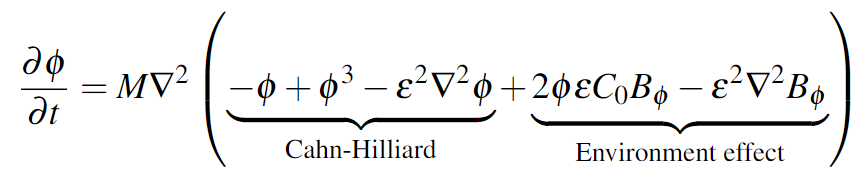
We match theory and experiment by scrutinising the influence of the coupling between the cells and the extracellular matrix. Our model simulates cell migration taking into account the extracellular matrix structure and the distribution of the biological factors.
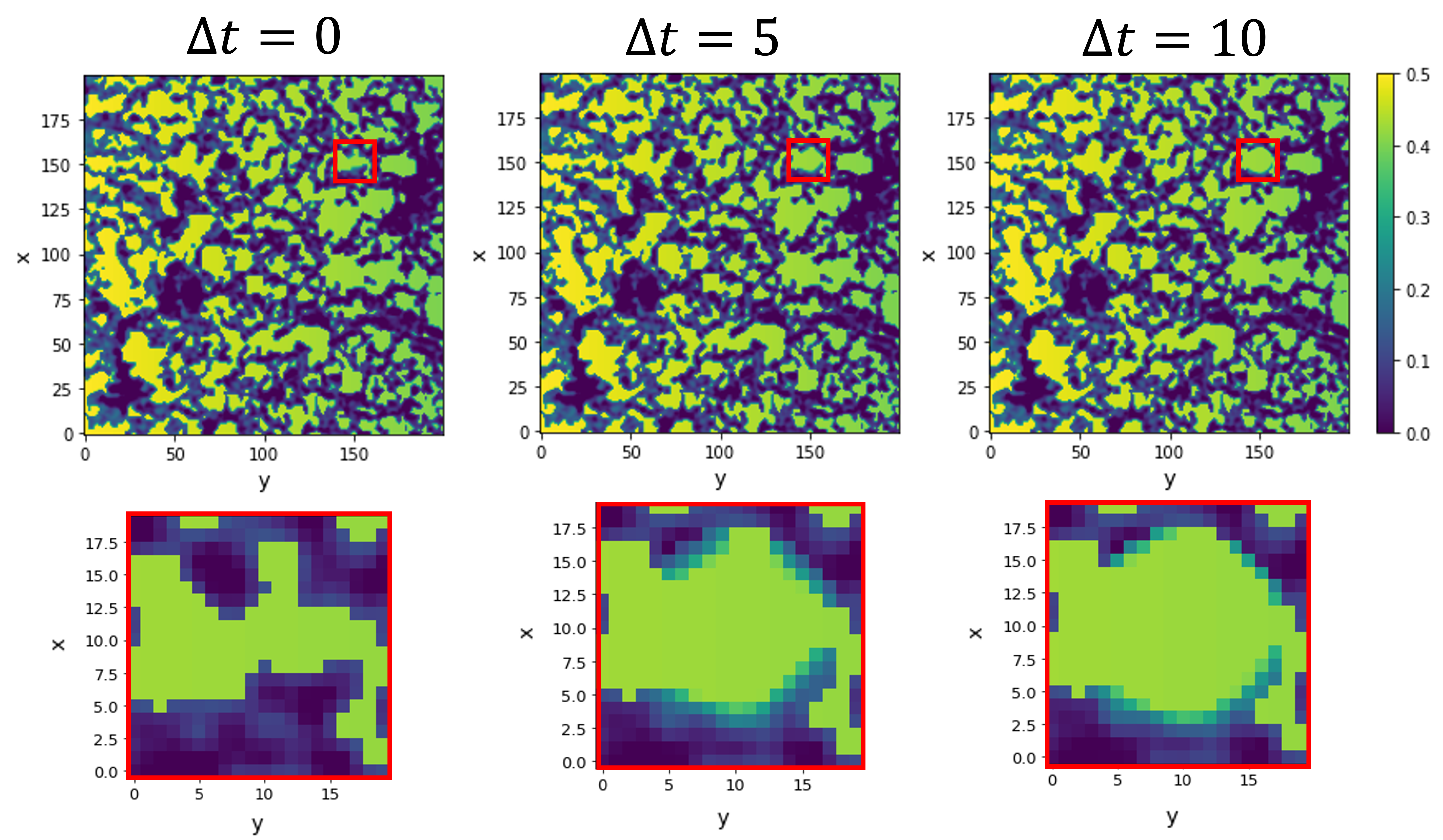
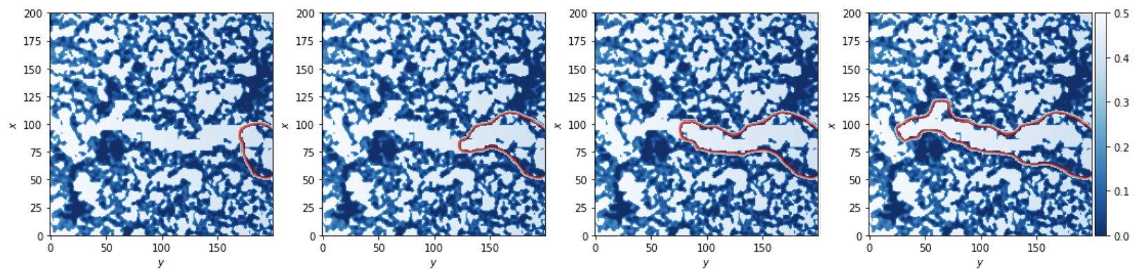
Our findings highlight the inherent nontrivial role of the extracellular structure in the sprouting-invasion dynamics and morphologies.
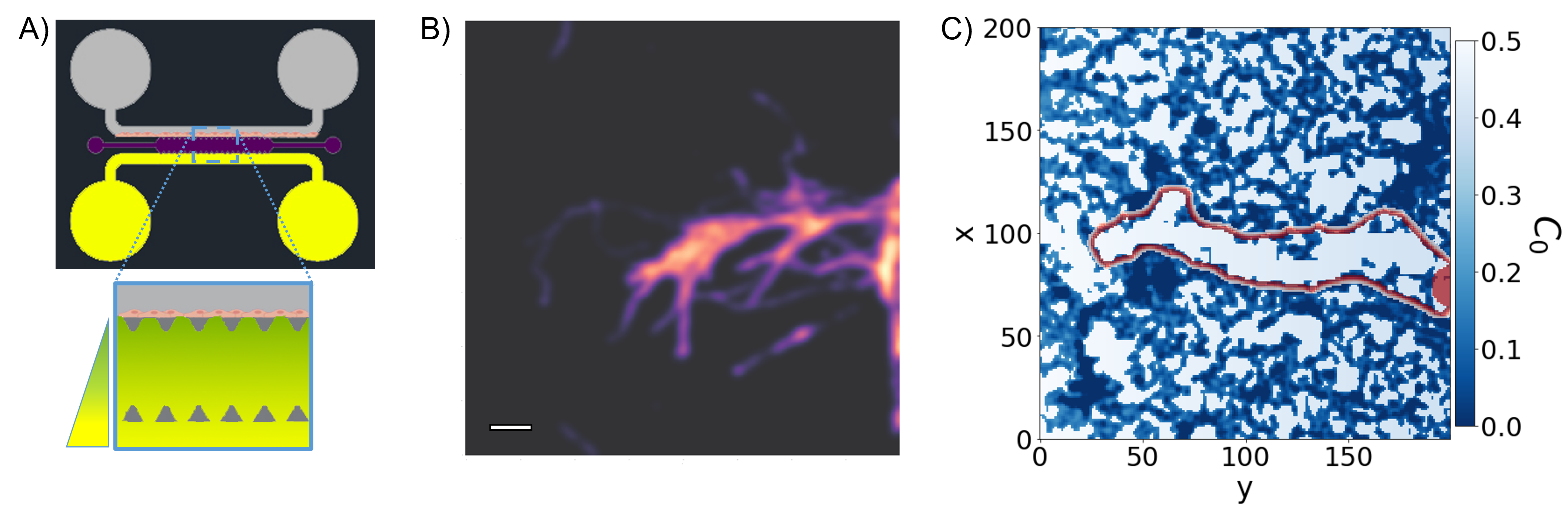
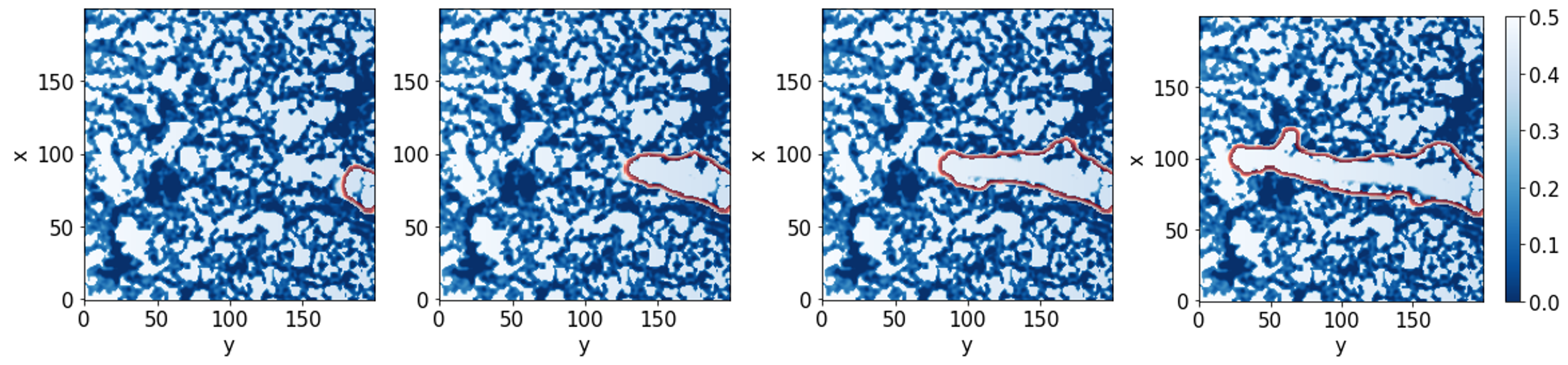
Panel A): experimental setup design. Panel B): cell advancement after 48h (the scale bar is 50μm). Panel C) is a sample of the simulation results.
Because of the strong interaction between cells and their environment, hydrogels have become of tremendous interest, given their biocompatibility, softness and water content. Used as an extracellular matrix, it provides a porous landscape for cells to migrate and invade. Moreover, given its water content, it is possible to solubilise biological factors to promote or inhibit cell invasion and differentiation.
The modelled cell environment is the digitalisation of a confocal microscopy image of the hydrogel (using NHS-rhodamine for fluorescence) plus an additional background of the distribution of the biological factors. The scale bar is 200μm.
In this project, we proposed a dynamic phase-field model to predict and describe the migration of a collection of cells due to chemotactic factors through the environment. Our formulation uses a single parameter (φ), and a partial differential equation describes the dynamics of the system:
Because the migration of ECs is oriented and regulated mechanically and chemically, we assume that the tip cell directs migration towards a clearer path in terms of extracellular matrix presence. Moreover, cells collective migration also involves the degradation of the extracellular matrix to enable the progression of cells. For this reason, we extended the model allowing tip cells to degrade the extracellular matrix by producing matrix metalloproteinases (MMPs).
Environment degradation for a static tip through multiple time steps (Δt)
Collective cell migration, with a small spherical reservoir and initial condition, through an open path with no extracellular matrix degradation.
The collective cell migration can sense the densest regions of the extracellular matrix and avoid them while breaking down and migrating through lower extracellular matrix dense regions in the system.
Object-oriented programming language with a huge ecosystem.
Image Processing (mask positioning) and Finite-element methods
Image processing for resizing and blurring image for modelisation.
If you want to know more about this work, extend it or collaborate
josep.ferre@fmc.ub.edu